Spine MRIQuick lookupPick the signal → get the type
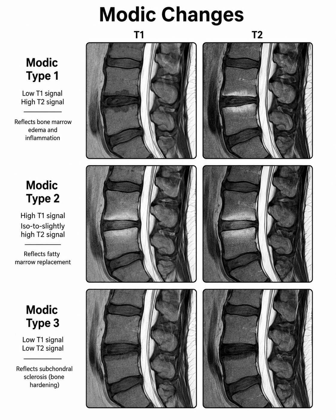
Modic Endplate Changes
Signal change in the vertebral endplate & subchondral marrow from disc degeneration. Tap the T1 and T2 signal you see — the type appears instantly.
What type is it?
T1 signal
T2 signal
Select a T1 and a T2 signal to identify the Modic type.
MRI reference
At a glance
| Type | T1 | T2 | Tissue | Key point |
|---|---|---|---|---|
| Type 1 | ↓ Dark | ↑ Bright | Edema / inflammation | Active; enhances; most likely symptomatic; mimics infection. |
| Type 2 | ↑ Bright | Iso–↑ | Fatty marrow | Most common; chronic/stable; suppresses on fat-sat. |
| Type 3 | ↓ Dark | ↓ Dark | Bony sclerosis | Matches dense endplate sclerosis on CT / X-ray. |
- Mnemonic: Type 1 = in1flammation (water: dark T1 / bright T2) · Type 2 = fat (bright T1) · Type 3 = bone (dark / dark).
- Where: most common at L4–L5 and L5–S1. Mixed types (1/2, 2/3) are common.
⚠️ Type 1 edema can mimic discitis–osteomyelitis. Modic spares the disc (low T2 disc, no disc-space enhancement or collection) — correlate clinically.
Reference only — not a substitute for clinical judgment. Classification after Modic et al., Radiology 1988.